Yesterday I reviewed several detailed architectural asymmetries between the right and left hemispheres, but presented little information on asymmetries in long-range connectivity. Recent advances in a form of magnetic resonance imaging called "diffusion tensor MRI" have made possible whole-brain imaging of white matter tracts, which are important for long-range connectivity in the brain. So, how has this technology refined the study of hemispheric structural asymmetry?

First, the basics: dtMRI analyzes the "fractional anisotropy" of water in tissue: in other words, it demonstrates the consistency of the alignment of water molecules within a single voxel (typically around 2 cubic milimeters). Interpretation of this is not easy, because anisotopy is influenced not only by myelination, but also water content, fiber diameter, and cell density. According to Buchel et al's 2004 Cerebral Cortex paper, water diffuses more quickly along the axis of an axon than it does perpendicular to that axon. Therefore, dtMRI will tend to trace out those axons that span large distances, such as those that are known to connect vastly separated regions of cortex.
Buchel et al.'s study demonstrated increased coherence in the left relative to the right arcuate fascicle, a bundle of white matter that connects the Wernicke's and Broca's areas, and is thus thought to be involved in language functions (i.e., myelination here increases with age, is generally lower in dyslexics and those with stuttering problems, and damage to this region results in aphasia). However, coherence was greater in the right inferior parietal lobe, thought to be involved in spatial processing.
In a 2004 Neuroimage article, Park et al. used dtMRI on 32 healthy adults, and found far more white matter asymmetries that Buchel et al. subjects. The corpus callosum had broadly greater coherence on the left than the right, as did other areas in lateral geniculate nucleus, cingulate, and cerebellum, whereas the uncinate fasciculus (connecting the anterior temporal and inferior frontal lobes) - among other regions - showed greater coherence on the right. Many other regions showed asymmetry but they were close to the midline; Buchel et al. suggested dtMRI may be prone to false positives for asymmetry in this region (although Park et al. used special algorithms that should have addressed this issue).
Although dtMRI & it's associated algorithms are still controversial, these studies are generally consistent with conclusions from other methodologies about hemispheric asymmetry in long-range connectivity: a tentative conclusion is that the left hemisphere has generally denser patterns of myelination (although there may be exceptions to this rule in the right superior parietal cortex). These results are also consistent with accounts of functional asymmetry, in which the left hemisphere is broadly more involved in language and the right more involved in spatial processes (at least for the right-handed).
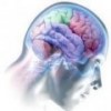
As the algorithms involved in dtMRI are explored more fully, it may be possible to draw conclusions from even more advanced techniques. For now, however, these methods are still being researched in the realm of applied computer science and mathematics. But the appeal is obvious: at the start of this article is the result of applying fiber tractography algorithms to diffusion tensor data.
Related Posts:
Asymmetric Architecture in the Left and Right Hemispheres
- Log in to post comments
