Today, we're going to look for rainbows in double-stranded DNA and see what they can tell us about DNA structure.
First, we're going to get a structure for a double-stranded molecule of DNA and open it in Cn3D.
If you want to do this at home and you haven't already downloaded a copy of Cn3D, you may want to read these instructions and get a copy. These directions also show how to download and open the structure. It's pretty simple once you've given it a try.
Hide a strand
Next, we're going to hide one of the strands. To do this, look in the menu bar for the Show/Hide menu and open it up. Choose Pick Structures.
A window will open up that shows a hierarchical list, with the individual structure first, the molecules that are part of that structure (in this case the two strands) and the domains that make up that structure (in this case, each strand has one domain).
Click the pointer somewhere inside the window to deselect the current structures, and then click either 1K9L_A or 1K9L_B. These are the names of the two strands.
Click the Apply and Done buttons.
We now pause briefly for a bit of background
Molecular viewing programs work by taking the three dimensional coordinates that show where elements are located in space, and drawing a picture or model. Some models are based on experimental data. Other models are hypothetical, they make predictions about what structures should be like by combining experimental data with structures that are predicted by modeling software. For example, some molecular modeling software will add the locations where hydrogens might be found. This is why I generally make a distinction between molecular viewing software - like Cn3D - and molecular modeling software (e.g. CaChe or ChemDraw).
Until recently, the PDB (Protein Data Bank) contained both experimental and hypothetical models, and the MMDB (Molecular Modeling Databank) at the NCBI, only contained models that were derived from experiments. If you want to know more about this, Deepak Singh has an interesting post on the recent change.
Okay, the mini-lecture is over, you can wake up.
Make it a rainbow
Because we're looking at drawings, created by Cn3D, from numerical data, we can ask Cn3D to do many different kinds of things with these drawings. We can use different drawing (rendering) styles, we can color molecules in different ways, lots of things!
We can even have Cn3D make rainbows!
To do this, we find the Style menu in the menu bar, click it, and choose Coloring Shortcuts.
And, we choose Rainbow.
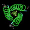
 You should see something like this image. Only one strand is showing and we have a rainbow coloring pattern, ROYGBIV, from one end (5' is red) to the other end (3'). The terms 5' and 3' refer to the structures at the different ends. We commonly use these identifiers (5' and 3') to tell one end from the other.
Okay, put your thinking caps on, it's time to learn something new.
Open up the Show/Hide menu and choose Show Everything to make the other strand reappear.
Look at the rainbow colors in your DNA molecule.
What does the coloring pattern tell you about the structure of double-stranded DNA?
TTFN!
technorati tags: DNA structure, digital biology, molecular modeling, Cn3D,
molecular structures
- Log in to post comments