
tags: researchblogging.org, dinosaurian soft tissue, fossils, bacterial biofilms, paleontology, endocasts, formerly pyritic framboids, collagen
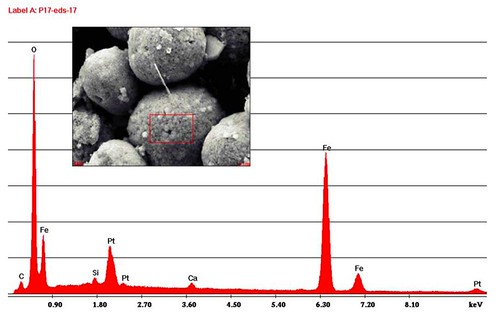
Figure 1. EDS spectrum of framboid. EDS spectrum of framboid showing an iron-oxygen signature. Pt is from coating for SEM. Area in red box was scanned for elements. [larger view].
![]()
Some of you might remember a paper published in Science that rocked the paleontological world by revealing that a broken thigh bone from Tyrannosaurus rex contained soft tissue. When this soft tissue was analyzed, it was identified as collagen from the blood vessels and, because it was found inside a dinosaur fossil, it was assumed to be ancient dinosaur tissue. Analysis of the protein sequence of this collagen indicated that chickens are the closest living relatives of this iconic dinosaur. However, a research team from my alma mater, the University of Washington in Seattle, just published a paper that questions whether this soft tissue really is preserved dinosaur tissue. After meticulous analysis, they hypothesize that this biological material might actually be a bacterial "biofilm" instead.
According to the 2007 research teams led by Mary Schweitzer from North Carolina State University in Raleigh, and John Asara of Harvard Medical School and Beth Israel Deaconess Medical Center in Boston, reconstructed amino acid sequences of a proteinaceaus substance found in a fractured Tyrannosaurus rex thigh bone bore striking similarities to collagen found in chickens, while several other protein strands were similar to amphibian collagen.
However, paleontologist Thomas Kaye of the University of Washington, and his research team contend that what was really inside the T. rex bone was not collagen from the dinosaur's blood vessels, but instead, it was a slimy biofilm produced by bacteria that lived within the spaces once occupied by blood vessels and cells.
A bacterial biofilm is a micro-ecosystem consisting mainly of a community of diverse types of bacteria that produce a polymeric matrix that sticks them to a living or inert surface. One familiar example of a biofilm is that layer of slime that grows on a person's teeth overnight.
Kaye compares this to what happens if you leave a pail of rainwater sitting in your backyard. Basically, after sitting for several weeks, the inner walls of the bucket feel slippery from the bacterial film that has grown on its inner walls. After time, this biofilm hardens through mineralization and takes on the shape of the surface it grew on. This is the same process that causes the formation of hardened deposits known as calculus on a person's teeth.
"We are not experts in the field," cautions Kaye. "We are not disagreeing with the fact that their instruments detected protein. We are offering an alternative explanation" for the source of this material.
When he began this work, Kaye's goal was to be the second person to find preserved dinosaur tissue, although he discovered something far different. In short, Kaye was doing what scientists often do: he was trying to duplicate Schweitzer's and Asara's findings. To do this, he went to the same formation where Schweitzer's sample came from, dug up a 65-million-year-old fossilized bone from a turtle (figure 2), cracked it open, and looked at what was inside using a Scanning Electron Microscope (SEM) (figure 3);
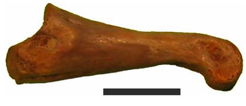
Figure 2. Well preserved complete bone used in initial investigation. Exceptionally well preserved small phalange from the Lance formation used for initial survey. No cracks or deformities present. Specimen was pressure fractured and directly examined under the SEM. UWBM 89327 Scale bar, 10 mm. [larger view].
DOI: 10.1371/journal.pone.0002808.
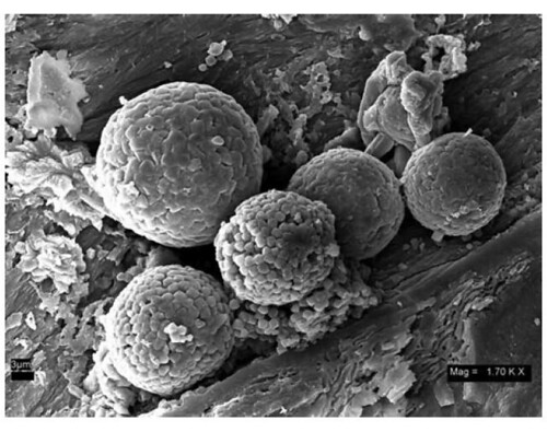
Figure 3. Iron oxide framboids. An iron oxide framboid cluster in dinosaur trabecular bone found commonly throughout time and taxa. At approximately 10 microns in diameter they are closely matched in size to red blood cells and typical pyrite framboids. UWBM 89327 Scale bar, 3 μm. [larger view].
Kaye's team was surprised when SEM revealed that these supposedly rare iron-containing structures were very common. What previously had been identified as fragments of blood cells due to the presence of iron were actually microscopic spheres containing iron, known as framboids.
The team found similar spheres in a variety of other fossils from different time periods. Interestingly, they also found iron-containing spheres in an ammonite fossil, a marine mollusc whose closest living relatives include the squid and octopus. These spheres were observed in a cavity within the ammonite fossil where blood had never been, so the residual iron could not have had any relationship to the presence of blood (figure 4A);
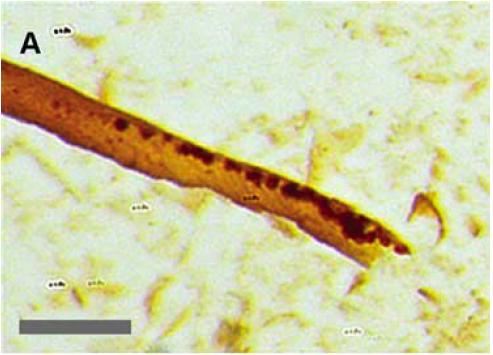
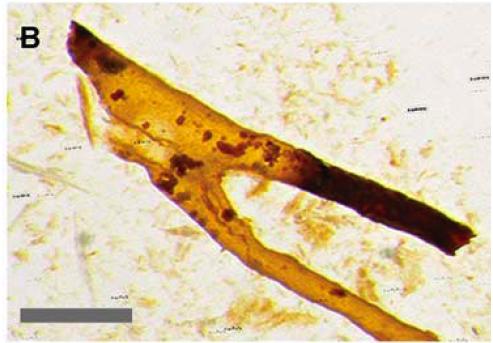

Figure 4. Tubular branching structures. Branching, transparent tube-like structures that match the porosity of the trabecular bone. Note small red grains that were found to be iron oxide framboids. These structures remain in acid baths after demineralization. Some are pliable, others frangible. Scale bars, 100 μm. Photos Z stacked, 7 images, unsharp mask, gamma adjusted.
After subjecting several fossils to demineralizing chemical baths as described in the original work, the team found a sediment consisting of a variety of branching structures that resemble capillaries (figure 4A-C). Some of these structures were soft and pliable while others were brittle and easily fractured, as described in Schweitzer's and Asara's research. These structures were then analyzed using energy dispersive spectroscopy (EDS).
Kaye's team then examined a variety of other bones, including a turtle's, and found similar structures. They also analyzed collagen and a modern biofilm that grew on microscope slides that they had placed into a nearby pond with a high iron content, and found that the fossil proteinaceous material more closely resembled slime from pond water (figure 9);
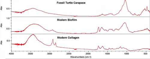
Figure 9. Infrared spectral comparison. Infrared spectra showing similarity of modern biofilms and modern collagen compared to fossil coatings. Cross correlation shows that the fossil material more closely resembles the modern biofilm than the modern collagen. [larger view].
The team also used SEM to examine some of the demineralized structures they recovered from the acid baths and found that they resemble decomposing bacteria
Rhodococcus sp. This species of bacteria has a variety of microscopic shapes, ranging from small, round cocci to long filaments (figure 10);
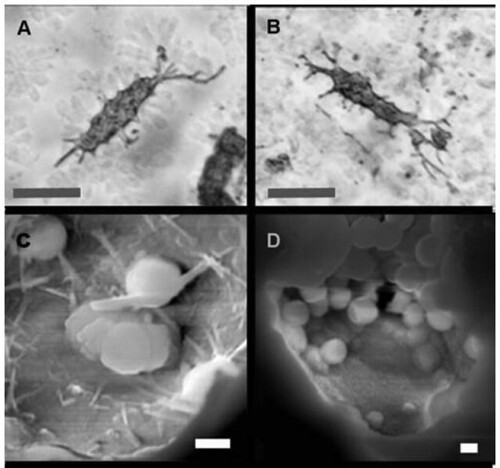

Figure 10. Osteocytes and lacunae. (A and B) Osteocytes found floating free in acid baths with fillapodia. (C,D,E) Fractured lacunae examined with SEM show filaments and spheres consistent with bacterial forms. UWBM 89325, UWBM 89322 Scale bars, A,B 10 μm, C-E 1 μm.
"We determined that these structures were too common to be exceptionally preserved tissue," observed Kaye. "We realized it couldn't be a one-time exceptional preservation," contrary to Schweitzer's and Asara's argument. This led Kaye's team to investigate the possibility that bacteria might be the source for this biological material.
As the result of their meticulous investigation, the team concluded that the voids in dinosaur bone provide the micro-environmental equivalent of a natural cave where bacteria can colonize and form biofilms. Further, when the cavity that biofilms have grown on is subsequently removed, as with an acid bath, the remaining biofilm retains much of the original morphology of the surface it originally grew on. This is the likely explanation for the quantity and similarity of structures found in fossil bone. This also indicates that these structures are unlikely to be preserved dinosaurian tissues but instead, are the product of common bacterial growth.
"From this evidence, we could determine that what had previously been reported as dinosaurian soft tissues were in fact biofilms, or slime," Kaye concluded.
Source
Kaye, T.G., Gaugler, G., Sawlowicz, Z., Stepanova, A. (2008). Dinosaurian Soft Tissues Interpreted as Bacterial Biofilms. PLoS ONE, 3(7), e2808. DOI: 10.1371/journal.pone.0002808.
Thanks - this means I don't have to struggle through the paper.
One up for the bacteria, I guess.
A possible logic:
1. Reptiles never stop growing during their lives. Old crocodiles becomes enormous.
2. Experiments have shown certain conditions cause piranhas to become giants.
3. These conditions were tuned to be similar to those before the Great Flood ~2300BC.
4. Reptiles in these conditions could have lived to be ancient, and therefore, enormous. Perhaps the size of dinosaurs.
5. These now extinct reptiles were enormous dinosaurs that went extinct sometime before the Great Flood, but not necessarily millions of years ago. Tissue from such a reptile could have survived to the present day within reason from such a dinosaur.
6. We make 'anything-into-oil' today using geological-scale pressures in an 85% efficient process at the Butterball Turkey plant in Missouri.
7. Carbon dating was advised against by its discoverer.
William - I see no logic whatsoever in your "idea", and no supportive evidence. You're quite evidently woefully ignorant about radiometric dating. Go read this:
http://www.asa3.org/ASA/RESOURCES/WIENS.html
Please, get yourself a good textbook about evolution. And then actually study it.
GS - This is rather sad. It would be nice to have some T.rex tissue, but OTOH, this does plainly refute creationist lies about evolutionists being a dogmatic clique. It's quite readily apparent how honest science is when we see papers published that question other papers.
It's remarkable - if this does indeed turn out to be bacterial biofilm - that the DNA most closely resembled chicken! I await with interest the next salvo in this investigation!
Uh oh...
I wonder where all the fossils are of smaller T. rex. By William's logic and some simple demography, one would expect there to be more smaller T. rex. in the fossil record, as there would have been more younger ones roaming the land chewing on coconuts.
william, it's too early in the day (and in the week) to be drinking while posting to blogs.
ian, i also wondered about the collagen sequences that the original team derived from the dinosaur bone -- i didn't know that bacteria produce collagen. i also didn't know that bacteria taste like chicken.
Ah, I thought I had seen something in the paper about collagen and bacteria:
Of course, that doesn't say that the bacterial sequences taste, err look like chicken collagen. More research required etc. etc.
The identification of collagen sequences in the T. rex has been challenged as well - I don't know enough to assess the arguments.
bob; yeah, that's the key phrase: RECENT discoveries. my knowledge of bacterial biochemical pathways and molecules is a few years old now. but "collagen-like molecules" leaves a lot to the imagination, especially when talking about specific amino acid sequences.
i assume that research of bacterial collagen-like molecules will become a more interesting field now that the broader implications are more widely appreciated.
But cephalopods have copper-based analogue to hemoglobin, and thus don't have iron in their blood anyway. So is whether the cavity was a place where blood had ever been even relevant?
i think they might mean that since ammonites could have eaten animals with iron-based blood, this is a possibility that cannot be excluded. since they specifically mention it, this must be important to note, although they don't explain why, so that's my guess.
very informative post..........Congrats.....
Hmmm...too bad.
Granting that what was originally found really is bacterial biofilm, is this then an obvious case of, "seeing what you want to see?" In other words, would researchers have identified the proteins as bird-like if they had just been blindly given a sample of it? Or was it like they knew it was a dinosaur bone, so they tailored their test to detect dinosaur proteins? If they would have thought it was from a fish fossil, for example, would they likely have discovered some kind of fish-LIKE protein (actually from bacteria)? Basically it seems strange to me that the biofilm they found 'just happened' to resemble dinosaur tissue. Of course that's if these new claims hold up.
William-
It's impossible to see where you're going with this.
"6. We make 'anything-into-oil' today using geological-scale pressures in an 85% efficient process at the Butterball Turkey plant in Missouri."
That's great, except before modern times, they didn't have Butterball Turkey plants.
My favorite part of Jurassic Park is when the girl breaks into the computer system. It makes no sense!
The problem here is the finding that the alleged Dinosaur DNA resembled Chicken DNA. If instead it was bacterial it seems to call the validity of DNA analysis itself into question.