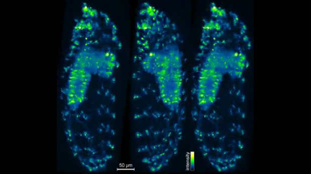
 A still shot from the 3D-IsoView microscope shows neural activity within a Drosophila larva detected with fluorescent indicators. Image courtesy of Keller Lab, HHMI/Janelia Research Campus
A new kind of three-dimensional technology, called IsoView, allows researchers to view biological processes within nontransparent animals that are rather large by microscopy standards such as the drosophila larva above and even the brains of larval zebrafish. According to the article in Nature Methods, the new type of microscope developed at the Janelia Research Campus (Ashburn, Virginia) gives high resolution images in each dimension captured at high speed, which is an improvement over prior microscope systems. Using the new technology, researchers will be able to image biological processes in a live animal in more detail that previous technologies allowed.
You can watch a sample recording of neural activity in drosophila here.
In a recent press release study author Philipp Keller stated: “We had decent microscopes for the type of imaging that we do—rapid imaging of cellular dynamics in large, living specimens. The temporal resolution matched the timescales of the processes we’re looking at, and we had microscopes that could give us good coverage and allow us to image for a long time without perturbing the system. But we hadn’t really tried to push spatial resolution much in our microscopes to date.”
Sources:
RK Chhetri, F Amat, Y Wan, B Hockendorf, WC Lemon, PJ Keller. Whole-animal functional and developmental imaging with isotropic spatial resolution. Nature Methods. 2015.doi:10.1038/nmeth.3632
- Log in to post comments