
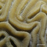
Scleractinian corals, also known as stony corals -- or just hard corals -- are the primary reef builders in the oceans. Their polyps secrete calcium carbonate to form a skeleton. A minority of species live as single polyps, but most stony coral species are colonial, and the structures they build 'grow' over time. They form a myriad of shapes: mounds, branches, fingers, plates, and encrustations.
In several previous posts I discussed and displayed photos of a number of so-called soft corals, all of which are octocorals, i.e., their polyps have eight tentacles. Stony corals are…
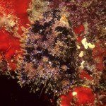
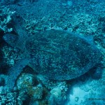
Many animals in the sea have evolved colors and forms that allow them to blend in with their surroundings. Some animals use their camouflage to hide from predators -- and some predators use camouflage to fool their prey.
It can be difficult to photograph such animals, partly because it's often hard to find them in the first place. If you look carefully at the photo at right, you will be able to make out the shape of a small purplish slipper lobster (Parribacus antarcticus), right in the center of the photo.
The picture was taken inside an underwater cave in Hawaii, and the lobster was on…

They look like a cross between a caterpillar and a tricked out centipede. They crawl about with considerable agility, they are voracious feeders, and they certainly know how to defend themselves. Meet the Bearded Fireworm (Hermodice carunculata), a free-moving marine Polychaete worm.
This species is widely distributed from the Caribbean, throughout the warmer waters of the Atlantic Ocean (including Florida and the Bahamas), and all the way to the Mediterranean Sea. All of the images in this post were captured in the Mediterranean, in the waters around Cyprus and Greece.
H. carunculata is…

The pretty creatures pictured here look like anemones, but they are not true anemones. They are Cerianthids, commonly referred to as 'tube anemones', which are taxonomically quite distinct from true anemones.
Cerianthids and true anemones do belong to the same phylum, Cnidaria, and the same class, Anthozoa, but tube anemones belong to the subclass Ceriantipatharia, a taxon that also includes the so-called 'black corals' (Antipatharia).
One of the visible features that distinguishes Cerianthid tube anemones from true anemones is the morphology of their tentacles. The macro photo below…
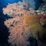
Sea fans are among the most beautiful sights seen by divers. Gorgonian sea fans are Cnidarians that build colonies in branching formations that usually are fan-shaped, thus the common name.
Like the Nephtheid soft corals I wrote about recently here on Photo Synthesis, Gorgonians are octocorals: each polyp has eight pinnate tentacles which it uses to capture nutrients suspended in the water column. They are seen most often on reef crests, or jutting out from drop-offs or steep banks in locations where natural currents will sweep plankton and other organic nutrients across the polyps'…
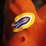
Nudibranchs -- marine snails without shells -- make wonderful photo subjects for the macro photographer. They are small, colorful, and they move slowly (as snails are wont to do). That last characteristic is particularly welcome. Most fishes are in motion almost constantly, and non-sessile invertebrates tend to scurry hither and thither. It's nice to find a subject that is not only photogenic, but doesn't turn tail or flat out disappear before the photographer can focus the camera's lens!
It's always interesting to find out and record which critters feed on what. Here are some macro…

Many animals in the ocean seek shelter from predators by living on or among other animals. Among fishes, members of the Damselfish family (Pomacentridae) often seek protection this way. Some of these relationships also are commensal or even symbiotic.
One of the most well known symbiotic relationships in the marine world is that between anemones and fishes commonly known as 'clownfish' or 'anemonefish'. Clownfish form a subfamily, Amphiprioninae, of the Dameselfish family. Each of the twenty-some species in this subfamily lives symbiotically with one or more anemone species.
Both the…

Everyone knows that some terrestrial animals are active primarily at night and sleep most of the day, while others go about their business during daylight hours and rest when it's dark. For some reason, many people are surprised to learn that the same thing holds true for animals that live in the sea.
One of the many marine animals that works the night shift is the crinoid species pictured here: Lampometra klunzingeri, a member of the Mariametridae family. During daylight hours, these crinoids hide in crevices in the reef. Shortly before sunset, like clockwork, they emerge from their…

Crinoids, a class of marine animals in the phylum Echinodermata, are pretty creatures. The photo at right shows a crinoid perched on a Malaysian reef with its fluffy arms extended for feeding. Looking at the photo, it's easy to see how they acquired their common name, Feather Stars.
This is how divers usually see crinoids, and this is how they are most often photographed. As pretty as crinoids are -- and they come in a vast array of colors -- photographs of the whole animal don't reveal much about the animal's structure or behavior. Whole-animal photos of creatures like this should be…
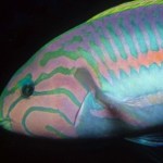
I like to photograph the faces of creatures that live in the sea. Here is a sampler of fish faces. All of these individuals belong to the Wrasse (Labridae) family. Most wrasses seem to have attractive markings on their faces, which show up well in close-up images like these.
Shown above: Thalassoma klunzingeri, from the Red Sea, about 15 cm (six inches) long. This one was photographed near Sharm el Sheikh, Egypt.
Shown above: Halichoeres hortulanus, about 23 cm (nine inches) long. Also from the Red Sea, this one was photographed at Ras Mohammed, at the tip of the Sinai Peninsula.…

Even the clearest water has at least some particles suspended in it -- sand, silt, plankton, who knows what -- and most of the time, in most places, the water isn't really all that clear. In fact, during a plankton bloom or after a storm that has caused a lot of runoff, you might just as well leave your camera on the boat. Light from a camera strobe has a nasty habit of bouncing back from any particulate matter that it encounters between the camera lens and the subject. The result is the bane of underwater photography: backscatter. Your images will look as though they were taken in a…

It is probably safe to say that when most people think of colorful things in the sea, tropical reef fishes come to mind first, followed perhaps by nudibranchs or sea stars. While most reef fishes, nudibranchs and sea stars are not only colorful but beautifully patterned, as a photographer, my favorites among colorful things in the sea are soft corals of the family Nephtheidae -- especially those of the genus Dendronephthya. They come in an incredible array of hues and color combinations, ranging from soft pastels to brilliant reds and golds.
Nephtheids are plentiful on tropical reefs…
We see this happen all the time here in Hawaii: Tourists go snorkeling -- sometimes for the first time in their lives -- and they are excited by what they see. They decide they want to take pictures of all the pretty fishies and corals to show their friends back home. They buy a single use waterproof camera, they snap away, and they are sorely disappointed when they see the result. Most of the photos are blurry, and though they thought they were shooting in color, all of the images are monochrome -- blue monochrome.
For quite a few reasons, taking photos underwater is very different from…

New month, new topic here on Photo Synthesis: underwater photography.
For the next four weeks I will be presenting photos of marine invertebrates and fishes from warm water locations around the world. These photos are the product of what has been, for me, a long journey of discovery -- about the sea, about marine life, and about photography.
My photos are documentary in nature, rather than works of art. That is not to say that I don't strive for a certain level of eye appeal, but my purpose always has been to record which creatures live where, what they look like in their natural…

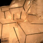
The lure of minerals is irrepressible when one sees fine crystals on display. This post is the first of four in a row featuring crystals seen at rock and gem shows in Arizona. The Tucson Rock and Gem Show is one of the oldest and largest of all such shows, with famed international reputation. The images shown in these posts display fine specimens on display and available from vendors. Most photos were taken with various point and shoot cameras using available light and no special preparation for the best photography conditions. Each image has a link where interested visitors to this blog…
Introducing a month long exploration of natural wonders seen by a woman who always has a camera on hand. We begin exploring with the wonders of minerals.
"Velvet Beauty" is a magnificent collector's specimen from Bisbee, Arizona. This museum-quality piece of malachite and azurite was available for purchase at a rock and gem show for a mere $25,000. See a higher resolution image here.
A titanium wash over a piece of quartz crystal is heat-treated to produce great color and flash. Seen on display with a vendor at the world-famous Quartzite, Arizona Rock and Gems show. Higher resolution…
As I prepare to hand off this photoblog to Cobalt123, I thought I would share my favorite non-rocket photos. Each clicks through to a story or geeky observation.
Last Thoughts
Magic Toes
Fire & Ice
A Beautiful Computation
in the Wolfram sense
Curiosity
Diamond Age & Eyes
and even some people
Namaste.
The Shuttle is glorious after midnight awaiting a big adventure...
A complicated design...
Lifts with a thunderous roar...
SRB Separation is visible to the naked eye... and the payload joins the ISS...
As the Shuttle is retiring from operation, SpaceX is gearing up to service the space station and other orbital launch needs:
Here you can see the music video of their first launch to orbit.
Alluring, enticing, begging us to come pay a visit...
Like a moth to the flame, before there were flames...
Reflecting our reflections...
and echoing in the dreams of Neil Armstrong...
For my last rocket-centric post, let me show the big project that I helped with, from a new prefecture for civilian space explorers, called Rocket Mavericks....
She lept off the pad with a glorious 30 ft. plume.
Computer simulations estimated she would scream to 100,000 ft. at Mach 3
⢠22 ft. tall, 540 lbs
⢠Booster stage: machined aluminum fin can and inter-stage coupler mounted directly on the Q motor casing.
⢠Upper stage: Custom wound composites, titanium tip
⢠Festooned with redundant computers, GPS, RF transmission
⢠Q booster staging to a P motor with a custom forward thermite…