microscopy
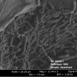
The Harvard Microbial Sciences Initiative Graduate Consortium hosted a fun workshop during the January term where students learned about microscopy by taking some amazing pictures of food microbes. The images taken with the scanning electron microscope of sauerkraut, kombucha, and some stinky cheeses show beautiful and complex landscapes made by and of microbes.
Sauerkraut is fermented by cute rod-shaped lactic acid bacteria:
Kombucha, a tea fermented by a combination of yeasts and bacteria, looks incredible under the microscope, where you can see the dense mesh of cellulose fibers that the…
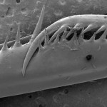
This looks like it could be painful. What is it?
Five points to the first person to name the organism, and five for the structure.
The cumulative points winner for the month of May will win either 1) any 8x10 print from my insect photo gallery, or 2) a guest blog post on the (safe-for-work) topic of their choosing.
[SEM image by the lovely and talented Elle Camino]
tags: Every Pollen Grain has a Story, pollen, microscopy, forensics, pollen signature, pollen fingerprint, science, Bosnian war crimes, pollinology, Jonathan Drori, TEDTalks, streaming video
Pollen goes unnoticed by most of us, except when hay fever strikes. But microscopes reveal it comes in stunning colors and shapes -- and travels remarkably well. Jonathan Drori gives an up-close glimpse of these fascinating flecks of plant courtship.
TEDTalks is a daily video podcast of the best talks and performances from the TED Conference, where the world's leading thinkers and doers give the talk of…
tags: The Nature of Cell Science, cell biology, microscopy, research, science, music, Venetian Snares, Szamár Madár, field work, Dirk Pacholsky, streaming video
Cell biologist, Dirk Pacholsky, created this video. He writes; "Certainly it´s irony in the title, because these images never made it to publication status. Sitting over the microscope, whilst listening to music the idea popped up to combine microscopic imagery with electronic music. The Scientist will certainly see more than a carpenter, but both might enjoy the beauty. Concerning the accompanying music, it is Venetian Snares (oh…
RESEARCHERS at the University of California, Berkeley have developed a microscope attachment which enables a standard mobile phone with a camera to be used for high-resolution clinical microscopy. Daniel Fletcher and his colleagues describe the CellScope in a paper published today in the open access journal PLoS One, and demonstrate that it can be used to capture high quality bright field images of the malaria parasite and sickle blood cells, as well as fluorescence images of cells infected with the bacterium that causes tuberculosis. The device could potentially become an important tool for…
A few years ago I needed to image some ants for a short taxonomic paper. Lacking a decent specimen imaging system (like Entovision), I decided to snap the photos at home using my standard macro gear: a dSLR with the Canon MP-E lens. The images turned out fine and were published in Zootaxa with the paper.
Later, the Antweb team imaged the same species using their standard set-up: a high-res video camera on a Leica microscope, focus-stacking the images with specialized software. I decided to compare the two. Here they are (click on each to view the uncompressed file…
It was a wet and rainy day yesterday, and we have a dissecting microscope, so I decided to see if I could find some tardigrades.
Tardigrade photo by nebarnix
Reposted from Nov. 2006
I went outside and scraped a bit of moss and some lichens off of our deck. Then I put the lichens and moss in a dish. We don't have distilled water in our house, so I added a bit of cool some tap water to the dish. I squeezed the moss and lichens in the water. Then I took a pipette and transferred a bit of the stuff in the water to a plastic petri dish and looked for tardigrades.
Sure enough, I saw one…
Heterospilus sp., head & compound eye, Costa Rica
Here are some shots from my training session this morning at the Beckman Institute's Scanning Electron Microscope (SEM). I haven't used SEM for years- wow! Great fun. Click on each image to enlarge.
Heterospilus sp. mesosoma
Heterospilus sp., ovipositor
For contrast, here's a photo of a wasp in the same genus taken with my standard Canon macro gear:
Heterospilus sp. Costa Rica, taken with a Canon 20D dSLR & macro lens
We'll be deciding over the coming months which type of images to use for our project.Â…