Cn3D
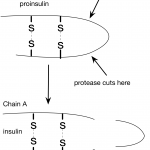
In my last post, I wrote about insulin and interesting features of the insulin structure. Some of the things I learned were really surprising. For example, I was surprised to learn how similar pig and human insulin are. I hadn't considered this before, but this made me wonder about the human insulin we used to give to one of our cats. How do cat and human insulin compare?
It turns out, that all vertebrates produce insulin, even frogs and zebra fish. Human preproinsulin is only 110 amino acids long and even human and fish insulin are pretty similar…
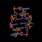
Today (4/25) is national DNA day. Digital World Biology™ is celebrating by sharing some of our favorite structures of DNA. We created these photos with Molecule World™ a new iPad app for viewing molecular structures.
As we are taught in school, the double stranded DNA molecule is a right-handed helix as determined by Watson and Crick using Franklin's x-ray diffraction images [1]. This B-form of DNA has approximately 10 nucleotides per turn of the helix and is the most common form of DNA found in nature.
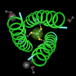
Classic structure with the elements colored.
Classic structure…
It's been interesting to watch as microbiology's own cold fusion debate has been raging. It began with an extraordinary claim about bacteria using arsenate as a replacement when phosphate concentrations are low (1).
It progressed when at least two scientist / bloggers ( here, and here) (not bloggers! the horrror! how uncivil!) gave public "journal club" presentations on blogs (envision dripping slime).
It continued with the science journalists lamenting about having swallowed the hype.
And it seemingly ended with another scientist / blogger's post that seemed to equate discussions…
Nick's post on Amantadine resistance in swine flu was so interesting, I had to look at the protein structures myself.
I couldn't find any structures with the S31N mutation that Nick discussed, but I did find some structures with the M2 protein and Amantadine. Not only are these structures beautiful, but you can look at them and see how the protein works and how the drug prevents the protein from functioning.
As Nick mentions, the M2 protein from influenza makes a channel for hydrogen ions within the viral membrane. The channel controls the pH inside the virus by opening and closing.…
Want to learn more about Parkinson's disease? See why a single nucleotide mutation messes up the function of a protein?
I have a short activity that uses Cn3D (a molecular viewing program from the NCBI) to look at a protein that seems to be involved in a rare form of Parkinson's disease and I could sure use beta testers.
If you'd like to do this, I need you to follow the directions below and afterwards, go to a web form and answer a few questions. Don't worry about getting the wrong answers. I won't know who you are, so I won't know if you answered anything wrong.
If you have any concerns…
Lots of bloggers in the DNA network have been busy these past few days writing about Google's co-founder Sergey Brin, his blog, his wife's company (23andme), and his mutation in the LRRK2 gene.
I was a little surprised to see that while other bloggers (here, here, here, and here) have been arguing about whether or not the mutation really increases the risk to the degree (20-80%) mentioned by Brin, no one has really looked into the structure and biochemistry of the LRRK2 protein to see if there's a biochemical explanation for Parkinson's risk. I guess that task is up to me.
Let's begin at…
Over 2600 genetic diseases have been found where a change in a single gene is linked to the disease. One of the questions we might ask is how those mutations change the shape and possibly the function of a protein?
If the structures of the mutant and wild type (normal) proteins have been solved, NCBI has a program called VAST that can be used to align those structures. I have an example here where you can see how a single amino acid change makes influenza resistant to Tamiflu®.
This 4 minute movie below shows how we can obtain those aligned structures from VAST and view them with Cn3D.…
In the class that I'm teaching, we found that several PCR products, amplified from the 16S ribosomal RNA genes from bacterial isolates, contain a mixed base in one or more positions.
We picked samples where the mixed bases were located in high quality regions of the sequence (Q >40), and determined that the mixed bases mostly likely come from different ribosomal RNA genes. Many species of bacteria have multiple copies of 16S ribosomal RNA genes and the copies can differ from each other within a single genome and between genomes.
Now, in one of our last projects we are determining where…
Have you ever wondered how to view and annotate molecular structures? At least digital versions?
It's surprisingly easy and lots of fun.
Here's a movie I made that demonstrates how you can use Cn3D, a free structure-viewing program from the NCBI. Luckily, Cn3D behaves almost the same way on both Windows and Mac OS X.
Introduction to Cn3D from Sandra Porter on Vimeo.
The grocery store magazine covers all say that home made gifts are big this year. So I thought, some of you might like to channel your inner Martha Stewart and make gifts with a science theme.
I'm here to help to you make a merry mug with one of our favorite molecules. Yep, we're talking caffeine.
1. First, we'll go to PubChem at the NCBI. It's not an exclusive (or even last) resort but there are lots of fancy molecules hanging around, just waiting to be discovered and put onto drinking containers.
2. Now, we'll look for a molecule. I'm going to use caffeine for this example since I…
Two protein structures from an avian influenza virus are shown below. One form of the protein makes influenza virus resistant to Oseltamivir (Tamiflu®)
Don't worry, these proteins aren't from H5N1, but they do come from a related influenza virus that also infects birds.
technorati tags: molecular models, protein structures, influenza, bioinformatics, Cn3D
One protein structure is from a strain that is sensitive to an anti-viral drug called "Tamiflu®". The other structure is from the same virus, except there's a slight difference. A single base change in the viral RNA changed the codon that…