Want to learn more about Parkinson's disease? See why a single nucleotide mutation messes up the function of a protein?
I have a short activity that uses Cn3D (a molecular viewing program from the NCBI) to look at a protein that seems to be involved in a rare form of Parkinson's disease and I could sure use beta testers.
If you'd like to do this, I need you to follow the directions below and afterwards, go to a web form and answer a few questions. Don't worry about getting the wrong answers. I won't know who you are, so I won't know if you answered anything wrong.
If you have any concerns or questions, please e-mail me: digitalbio at gmail dot com
My goal is to find out if doing the activities helps you understand (or think you understand) how a certain mutation might change the activity of a protein.
Background information
Parkinson's disease has been a big topic in the news lately with the announcement that Sergey Brin, co-founder of Google, has a form of the LRRK2 gene that increases his risk of developing the disease. The disease-related version of LRRK2, however; is only one of many genetic changes that can lead to the symptoms of Parkinson's.
In this activity, we will explore a different mutation that is also associated with Parkinson's disease and see if we can understand why this other mutation could lead to problems.
A mutation in ADH1C related to Parkinson's Disease
Alcohol dehydrogenases are enzymes that help protect the body from a number of toxic substances, including ethanol. Recently, an international study of several hundred patients identified a mutation in one of these genes, ADH1C, that significantly increases the risk of developing Parkinson's disease.
ADH1C encodes a member of the alcohol dehydrogenase family of enzymes called "gamma 2 alcohol dehydrogenase." This mutation changes one of the bases in the 78th codon of the ADH1C mRNA. Instead specifying glycine, the altered codon tells the translation machinery to stop adding amino acids to the protein chain. When the ribosome hits this codon, protein synthesis stops.
Your goal is to use Cn3D to view the normal ADH1C protein, annotate the structure to see how the mutation changes the protein, and last, explain what the mutation might be doing to the function of the protein and why the mutation might have that effect.
Instructions:
A. Get the Cn3D program and structure from the NCBI.
- Download and install Cn3D. **If you're using VISTA, get Cn3D here. Cn3D is part of the CDTree 3.1 package.
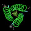
- Open 1HT0 from the NCBI. (Clicking the link should open the structure in another web browser window).
- Click the button below the image to download the structure.
- Open this structure in Cn3D.
B. Find where the two subunits interact
- Begin your investigation by finding some of the features in the protein. Open the Style menu and change the coloring style to "molecule" to see how the two chains fit together.
- Change the coloring style to "rainbow" to see which region of each chain is involved in holding the two chains together.
- Use your mouse to select and highlight the amino acids that you think are involved in holding the chains together. Adjust your selection until the amino acids that contact the other subunit are the only amino acids that are selected. Look at the numbers in the bottom left hand corner of the Sequence/Alignment window to identify where these amino acids are located within the protein. You may want to write this information down for later.
- a. Change coloring style to molecule
- b. Click the Sequence/Alignment Viewer window.
- c. Open the Mouse Mode menu and choose "Select Rows."
- d. Click one of the sequences in the Sequence/Alignment Viewer to select it.
- e. Click the structure window to get the structure menu.
- f. Open the Show/Hide menu and choose Select by Distance and Other Molecules. Leave the distance setting at 5 angstroms.
- g. Now, all the residues in other subunit that contact the one you selected, will be yellow. You can scroll through the Sequence/Alignment Viewer Window to see where they are.
Here is an alternative way to identify the amino acids that help hold the two subunits together:
C. Find the cofactor, the active site, and identify some critical amino acids in the active site
- Now, take a look at one of the subunits and find both of the zinc ions. One zinc ion is bound towards the outside of the subunit. The other zinc is bound near the cofactor, in the active site of the protein. The active site is where enzymes carry out chemistry. They break or join other molecules together and all of this chemistry happens at the active site.
- Use your pointer to click the zinc that's bound at the active site. When the zinc is selected it will turn yellow. In these next steps, we're going to identify the amino acids that are bound to this zinc.
- Open the Show/Hide menu and choose Select by Distance, then Other Molecules. You can leave the distance setting at 5 angstroms.
- Open the Style menu and select Annotate.
- Click the New button. This will make another small window pop up. In the small window, click Edit Style.
- Now, a panel will appear with several choices for changing the way that the residues are colored or drawn.
- a. In the first column of the panel, click the check box in the Protein sidechains row. Now, the amino acid side chains will be visible.
- b. In the Rendering column, choose Ball and Stick in the protein sidechains row.
- c. Click the Done button at the bottom of this panel.
- d. Click the OK button on the small Edit Annotations panel.
- e. Click Done in the User Annotations panel. At this point, the amino acids, cofactor and zinc should still be highlighted.
- Open the Show/Hide menu and choose Show Selected Residues.
- Zoom in to look at the zinc more closely. You'll see white colored bonds between the zinc and some of the amino acids. At this point, only zinc, the cofactor, and nearby amino acids will be highlighted. Click those amino acids, one by one and look for the highlighted letters in the Sequence/Alignment Viewer window to see which amino acids they are and where they're located.
Write down the names of those amino acids and their positions. Those amino acids are important in metabolizing toxic compounds, like ethanol.
The selected residues and the cofactor will be highlighted yellow.
Click your pointer somewhere inside the Sequence/Alignment Viewer to deselect the selected residues so they're no longer all yellow. If you're using Cn3D 4.2, you will need to deselect each amino acid indvidually by clicking it.
D. See how the mutation at codon 78 affects the protein
- Now,we're going to see what happens when the 78th codon contains the mutation we discussed earlier. Open the Show/Hide menu again and choose Show Everything. Both of the subunits will reappear.
- Find the 78th residue in the Sequence/Alignment Viewer (this is where the stop mutation occurs) and use your mouse to select ALL the residues in BOTH of the amino acid chains from number 78 to the very end of the protein.
- Open the Style menu and choose Annotate.
- Click the New button. The Edit Annotation window will appear as before.
- In the small Edit Annotation window, click Edit Style.
- In the first column of the Style Options window, change the Protein backbone option to "none" and make sure the protein sidechains box is unchecked.
- Click Done.
- In the small Edit Annotation window, click OK.
- In the User Annotations window, click Done.
- At this point, all the amino acids that would normally have been added after glycine 78, are hidden and you can see how the protein would be changed in the absence of those residues.
What do you think the loss of the residues after glycine 78 would do to the function of the protein? Why would the loss of those residues affect the function?
When you're done thinking about this, please go answer these questions.
Thanks!!
Thanks for giving me a reason to play with Cn3D. You have designed a nice exercise which might be helpful in explaining how single mutations have a large affect on a biological system.
Great idea! I'm pretty digital, but not very biological, so I hope you're not in a hurry for responses.
Thanks
I would like to adapt this to my NGBW site as an example.
Do you ave any objections?
Mark
Thanks for asking Mark!
I don't have any objections as long as I'm credited as an author.
Hi Sandra,
This is a nice activity. I do professional development for MS and HS teachers. I will have them do this activity tomorrow but I have some comments for you today:
Step 6: it is not readily apparent how the "rainbow" shows areas of chain contact.
Step 15: the white bonds are not seen until you click on the a.a. and turn it blue.
Step 17: I could not highlight and scroll past to window size or on both chains, is there a trick to doing this?
Thanks Simona,
That would be great! Please make sure to have them answer the questions in the form when they're done, that would be very helpful for me. The link is at the end of the activity, also.
I have some answers for your questions:
Step 6: The rainbow doesn't show where the two chains contact each other. The previous step where you color by molecule shows where the two chains are closest together.
I suggested using the rainbow coloring next in order to identify the relative locations of the amino acids within each chains. f you were to look at the structure and see that the amino acids in the contacting region are green for instance, you could look at the sequence and see that those green letters are between residues .... and residues...
I'll edit the activity, and add an alternative method for seeing contact points that I think might be more clear.
Step 15: Yes, I see what you mean. You don't see the white bonds until you've deselected everything (otherwise, everything is yellow). Okay, I'll clarify that instruction.
Step 17 Yes, there's a little bit of a trick. You need to use your mouse to drag and select a rectangular area from both chains, then let the mouse button go and scroll to the end of the sequence.
Press the Shift key down and keep it down.
Then, use your mouse to drag and select the remainder of the sequence for both of the chains.
This is a bit tricky when you don't have a mouse but I've done it on a laptop, it's possible.
Thanks for your comments!