In the class that I'm teaching, we found that several PCR products, amplified from the 16S ribosomal RNA genes from bacterial isolates, contain a mixed base in one or more positions.
We picked samples where the mixed bases were located in high quality regions of the sequence (Q >40), and determined that the mixed bases mostly likely come from different ribosomal RNA genes. Many species of bacteria have multiple copies of 16S ribosomal RNA genes and the copies can differ from each other within a single genome and between genomes.
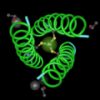
Now, in one of our last projects we are determining where the polymorphic bases map within the structure of the 30S ribosomal subunit (see a video for background information on ribosomes here).
This video shows how we align the sequences and find the polymorphic sites in the three dimensional structure.
We have two questions that we want to answer:
1. Do any of these polymorphic bases map within binding sites for an antibiotic? We're looking at tetracycline in this structure.
2. Do any of these polymorphic bases map within regions of secondary structure or in positions where the RNA is bound to a ribosomal protein? I think they wouldn't because of evolutionary constraints, but it's still interesting to find out whether this is true or not.
The video is about 15 minutes.
Mapping polymorphisms in 16S ribosomal RNA from Sandra Porter on Vimeo.

... and determined that the mixed bases mostly likely come from different ribosomal RNA genes.
Have you discussed the potential for chimeric sequences? It's a continual problem, as demonstrates.
Part of the reason I like working with the Nitrosomonadaceae is that they have only one rRNA operon. ;)
The rRNDB is a great resource. Actu
Thanks TomJoe,
I hadn't thought about that. It's easy to see how that would happen, unless people use physical methods like optical mapping or something to validate their assemblies.