What's in a picture?
Prof. Benny Shilo knows the value of a good picture. We recently mentioned his book: Life’s Blueprint, which uses photographs of things like bread dough and yeast cells to illustrate the process of biological development. Here is the image from the most recent piece we have uploaded on his research:
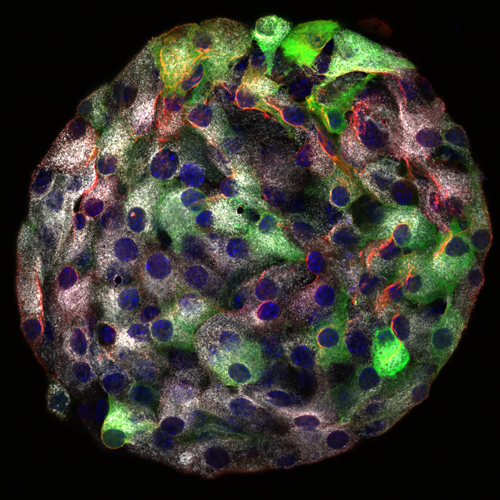
This is an individual Islet of Langerhans, as you’ve never seen it before. The white dots are the insulin-containing vesicles inside the beta cells, which both sense glucose levels and secrete insulin. Shilo and his team managed to get “close-up shots” of the individual cell membranes, and found that they have straight edges where both sensing and secretion functions are located.
We love the images in this new article on the work of Prof. Talila Volk because, aside from their eye-catching colors, these ones really do illustrate the story her work tells.
Volk’s work investigates how muscle fibers get renewed through exercise. It may all come down to a protein that senses muscle contraction and tells the DNA in the cell nucleus to make more muscle proteins.
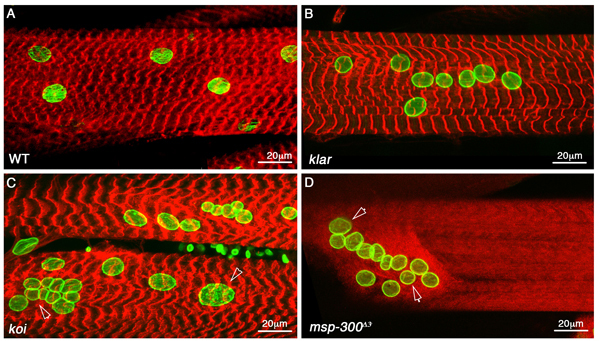
In this image, red is muscle fiber in fruit fly larvae, green is nuclei (muscle fibers are large cells with multiple nuclei) (A) shows normal muscle fiber, the others show what happens when the protein is missing.
How does it work? This image says it all:
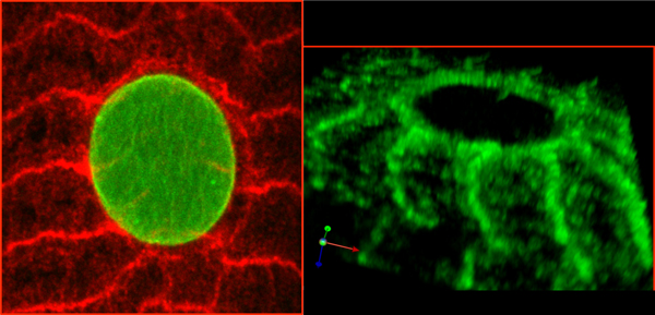
In the left image you can see how the protein structure encircles the cell nucleus. At the other end of its arms (red) it connects to the cell’s skeletal structure – the cytoskeleton. On the right, in green, you can see the protein structure. Its arms are springy – so that a pull on the end transmits a signal to the middle.
Volk and her group refer to the protein as a biological “mechanosensor,” and, indeed, there is something rather mechanical about the right-hand image.
Assuming that form follows function, Volk has surmised that similarly-shaped proteins in human muscle fibers do the same thing.
Third, we have some cancer research images from the lab of Prof. Lea Eisenbach:
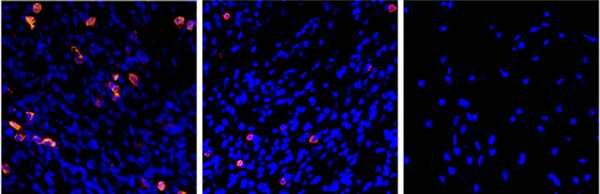
These tell the story of anti-cancer immune activity. Tissue and tumor cells appear in blue, pink shows immune cells that attack the cancer cells. When tumor cells appear in the back, there are many immune cells (left); but in the brain (middle) the same tumor cells attract relatively few immune cells. In fact, the normal brain tissue (in the same brain) on the right has no immune cells. If you first inject the tumor into the rat’s back, where there is immune activity, and then inject the tumor cells from the back into the brain, the brain will be protected from the cancer.
And, oh yes, they are pretty too. This image, again from Talila Volk, of a 3-D computer model of a fruit fly larva muscle fiber was the inspiration for a sewing project:

