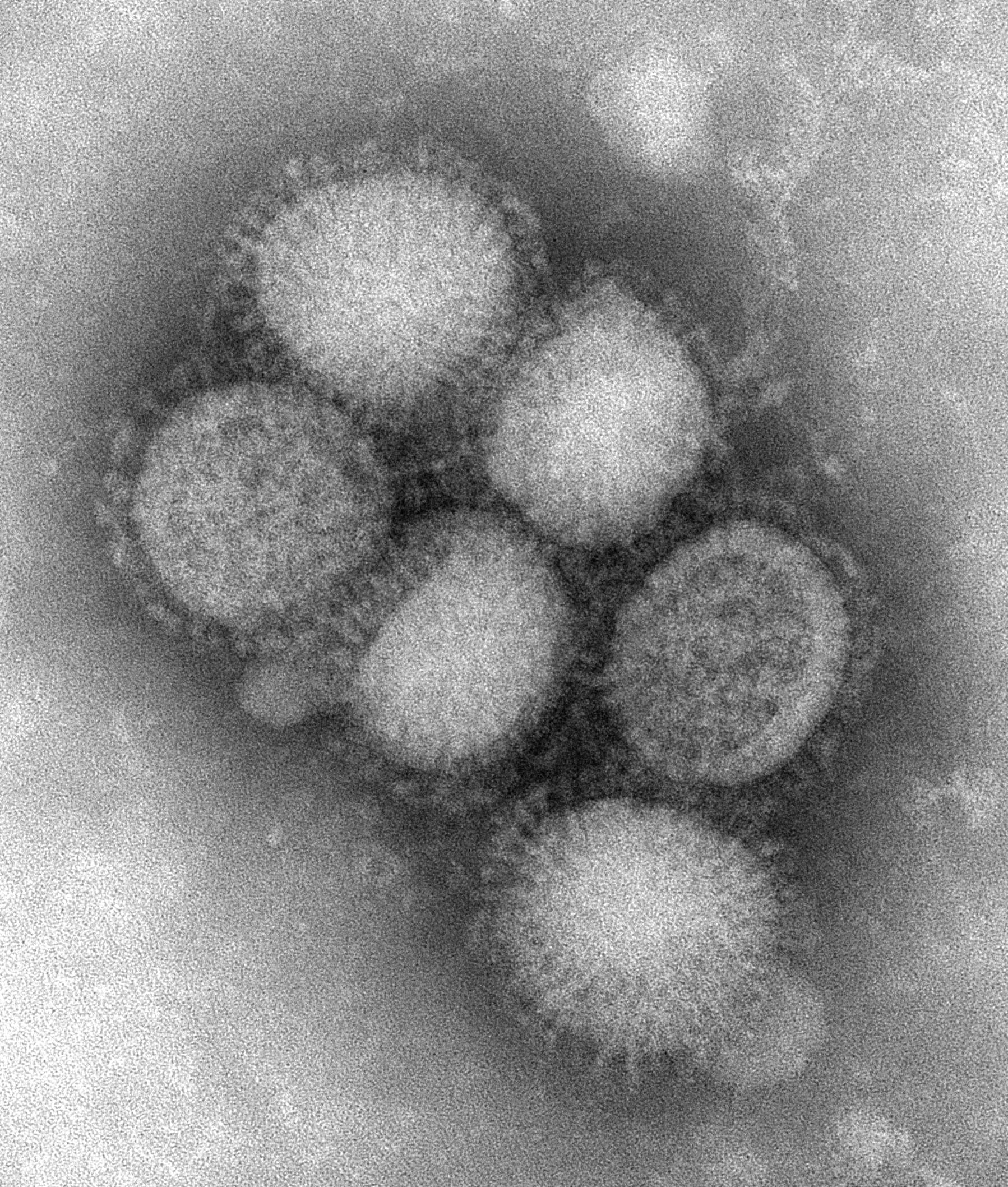
We talk so much about the flu virus we thought we'd show you some nice pics that CDC has just put up. This is a review for many of you put reviews are always helpful. In these three pics, only one is the actual swine flu virus, the other two being "cartoon" depictions of a generic influenza virus. The cartoons are quite nice and helpful to see what you are looking at in the electron micrograph of influenza virions (virus particles), probably grown in tissue culture. I say "probably" because there is no other information on the site other than the micrograph was taken in the CDC Influenza Laboratory, but when the virus grows in your lungs it usually isn't nice and spherical like this but assumes many shapes, often elongated and strand-like. Looking at the photomicrograph, though, you see the essentials. Here it is:

Large version here
There are six virus particles, spherical in shape, with a denser shell surrounding an interior space and a rough, brushy looking surface. Those brushy things are the surface proteins hemagglutinin (HA) and neuraminidase (NA). There are 16 broad kinds of HA (each with myriad lesser variations) and 9 kinds of NA (also each having lots of minor variations). That gives a total of 144 different broad combinations (16 times 9). The different kinds of HA and NA are unimaginatively named: just numbered H1 to H16 and N1 to N9. Any particular viral particle has only one of the 16 possible kinds of HA (called a subtype) and only one of the 9 possible kinds of NA and the particular combination gives the familiar subtype naming system, e.g., H1N1 or H3N2. Only a few of the combinations are found on flu viruses that infect humans. Most of the other subtype combinations are found in aquatic birds, the main reservoir for influenza viruses, but we know a number of other animals can be infected with one or another influenza virus, including pigs, cats, dogs and horses.
The two surface proteins (HA and NA) are usually portrayed as spikes sticking through a lipid bilayer membrane derived from the host cell that the virus dragged with it when its progeny budded off after infecting it. If you want to know more about lipid bilayers and the glycoproteins like HA and NA that stick through them we explained and illustrated a lot of it in a series of posts on the old site which we reprised in posts which start here. There are links to the others in the first one. But let's just stay with the pics for now and you can go back and fill in the details later.
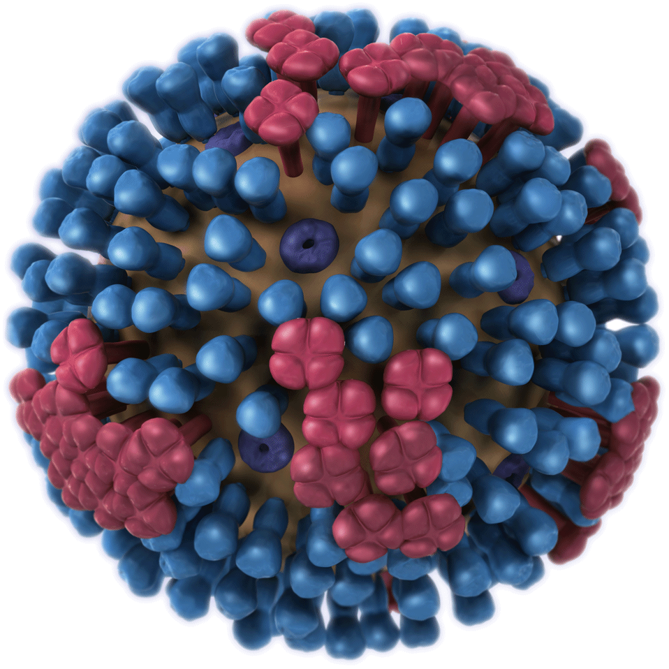
The first cartoon shows the viral particle from the outside, the HA (hemagglutinin) spikes in blue and the NA (neuriminidase) spikes in red (I had to reduce the size to fit on the blog page, but if you click on "larger version" you'll see each more clearly):

Large version here
The spikes are sticking through the stolen host cell lipid membrane (shown as a brown base, now called an envelope; viruses that do this are called enveloped viruses, so influenza virus is an enveloped virus). The spikes are attached under it to a hard shell protein, designated M1. M1 gives the virus a more rigid structure. Embedded in and studding M1 and also sticking through the membrane is another protein called M2 or the ion channel protein. You can't see it in the photomicrograph but it is shown in the two cartoon pics. There are more HA than NA on the surface and not much M2 visible. You'll see two M2s looking like little buttons, one above and one below the equator towards the left. The brownish stuff is lipid bilayer and you can't see the M1 protein on the first cartoon. The virus has genes that encode for HA, NA, M1 and M2 but not for the lipid bilayer which is scavenged from the host cell.
The HA and NA are involved with attaching to and entering the cell and again when the progeny leave it (more details here and here). The M2 ion channel protein is nicely explained by Vincent Rancaniello over at the Virology Blog, so head on over there for the details (it is involved in the antiviral adamantane class of drugs). You can get great explanations of many of the same things I'm talking about here (and much more) on the Virology Blog, written by a real virologist (not an epidemiologist). But we figure it's always helpful to have more than one explanation, so that's our excuse. That and the fact that we find it interesting and like to write about it.
So where are the viral genes whose sequences everyone talks about? They are in the interior of the shell. The second cartoon shows a cutaway view of the virus.

Large version here
Inside you see the eight genetic segments, labeled RNP. RNP stands for ribonucleoprotein, although the picture includes not only the protein but the RNA. So the squiggles are the virus's RNA (its genetic blueprint) packaged structurally with a viral protein. Not shown is the polymerase complex, three proteins that work together to replicate the long strands of viral RNA. These three RNA-dependent replication machinery proteins are called PA, PB1 and PB2. They are also in the interior of the virus. More here.
Clearly there is a great deal more to the virus than this description of its physical landmarks. But the pics are nifty and it's usually nice to put a face with a name, as they say. Reminder: The Virology Blog is a great place for details and recent news about the molecular biology. Highly recommended.
This is such a useful look at the virus, and a good explanation. I appreciate it. These pictures are great, but always remind me of the current problems with high resolution imaging, which is that you cannot see sub visible wavelength (nanometer scale) objects properly in motion. I am an Applied Physicist who usually deals with fixed objects. It seems to me, that following the movement patterns of viruses are equally important to seeing the morphology. This is an interesting challenge, and if it can be solved will lead to physical solutions to medical problems like this one. Of course this is my biased and perhaps naive outlook on the situation.
Matthew: Glad you find the pics helpful. I'm not exactly sure what you mean by the movement patterns of viruses. Viruses aren't alive by the usual standards. They don't eat, grow or move by themselves. They are passive in that regard. They are just a lump of protein and genetic material (and some lipid for enveloped viruses). There is a complex dynamic when the HA "finds" (by random encounter) a lock and key match on a host cell (the viral receptor). That sets in motion biological actions by the host cell which allow the virus to get inside the cell and hijack the protein synthesis machinery of the host cell. There are some nice animations on the net of some of this.
Then there's this representation of swine flu, which makes it look quite cuddly!
http://www.giantmicrobes.com/us/products/swineflu.html
A great gift for the epidemiologist in your life.
Here's a very cool video animation of a flu virus invading your body, courtesy of NPR
http://www.npr.org/templates/story/story.php?storyId=114075029
Thank you for the clarification. I tend to think rather abstractly about mption. For example I work in the field of rheology, which is macromolecular flow. I speak of molecules in a polymer when there is shear as I do here in refernece to virus . So when I say that we need to see a virus in action, I mean to know how it responds at different stages. I always tell students the shortcoming of AFM for example by saying that for complex systems you need a snapshot, and the differential. The same is true for biological systems, I think. I am of course showing plenty of ignorance on this, and your site is helpful.
Matthew: Ahh, I see your take on it now. It is even more complicated from that point of view because the virus is pleomorphic. The matrix protein isn't so rigid, the virion (the virus particle) has a lipid bilayer surface over it that can itself flow, and the whole thing can assume many different shapes (frequently filimentous) when in situ. Those shapes are the result of convective forces and movement is subject to the rheology you study, and at that scale, brownian motion as well. Then there are the interactions with large biomolecules like the receptors, involving van der Waals, hydrogen bonding and other electrostatic forces. Quite a mix.
Thank you Revere. This is an interesting rheological and nanocomposite issue in one. Thanks for filling me in. Now I want to get my hand on a specimen. What is the size of the virus? I have worked a little on condoms, and initially there was an issue with elastomer protection against anything in the 100 to 200 nanometers. Now that isnt a problem, because of fairly simple elatomer engineering. In my lab we are now working with graphine, (in situ, and in a soft polymer matrix), which can trap carbon and silica aggregates of this size. The issue being that Vander Walls are actually too easy to disagglomerate, making this graphine net difficult to control. Still I have this fantasy that a nanofiller like graphine could in fact be useful in neutralizing a virus. Since, as I have said before here, I have no idea what I am talking about as far as viruses, this may be naive. I just enjoy hearing you speaking of bonds, forces and material matrix. The language I use. Thanks again, and I look forward to reading and learning more.
Matthew: Since they are pleomorphic it is hard to give a size, but the minimum (spherical shape) is about 120 nm.